Tumor Vs Cyst On Mri -

Last update images today Tumor Vs Cyst On Mri

tumor vs cyst on mri        
        <figure class= Brain Mri Tumor   Curroncol 29 00590 G001
Brain Mri Tumor Curroncol 29 00590 G001
Brain Tumor Color Mri   Applsci 10 01999 G001
Brain Tumor Color Mri Applsci 10 01999 G001
Top 92 Pictures Pictures Of Brain Tumors In Adults Latest   MRI Brain Depicting A Solid Cystic Tumor At The Left Temporo Parietal Region The Cystic
Top 92 Pictures Pictures Of Brain Tumors In Adults Latest MRI Brain Depicting A Solid Cystic Tumor At The Left Temporo Parietal Region The Cystic
Cat Scan Brain Tumor   Fneur 13 932219 G001
Cat Scan Brain Tumor Fneur 13 932219 G001
MRI Scans Showing The Appearance Of Tumour Deposits Arrows Download   MRI Scans Showing The Appearance Of Tumour Deposits Arrows
MRI Scans Showing The Appearance Of Tumour Deposits Arrows Download MRI Scans Showing The Appearance Of Tumour Deposits Arrows
A C MRIs Showing A Cystic Solid Tumor MRI Magnetic Resonance   A C MRIs Showing A Cystic Solid Tumor MRI Magnetic Resonance Imaging
A C MRIs Showing A Cystic Solid Tumor MRI Magnetic Resonance A C MRIs Showing A Cystic Solid Tumor MRI Magnetic Resonance Imaging
MRI T1 With Gd Before Operation Showed A Round Enhancing Lesion Long   MRI T1 With Gd Before Operation Showed A Round Enhancing Lesion Long Arrow With A Cyst
MRI T1 With Gd Before Operation Showed A Round Enhancing Lesion Long MRI T1 With Gd Before Operation Showed A Round Enhancing Lesion Long Arrow With A Cyst
Postoperative MRI Resolved Tumor Completely Excised And Cyst Which   Postoperative MRI Resolved Tumor Completely Excised And Cyst Which Disappeared With.ppm
Postoperative MRI Resolved Tumor Completely Excised And Cyst Which Postoperative MRI Resolved Tumor Completely Excised And Cyst Which Disappeared With.ppm
A Axial T1 Post Contrast MRI Showing A 50 Mm Cystic Mass With A Solid   A Axial T1 Post Contrast MRI Showing A 50 Mm Cystic Mass With A Solid Intracystic
A Axial T1 Post Contrast MRI Showing A 50 Mm Cystic Mass With A Solid A Axial T1 Post Contrast MRI Showing A 50 Mm Cystic Mass With A Solid Intracystic
MRI The Tumor Contained Small Cysts Arrow Download Scientific Diagram   MRI The Tumor Contained Small Cysts Arrow Q320
MRI The Tumor Contained Small Cysts Arrow Download Scientific Diagram MRI The Tumor Contained Small Cysts Arrow Q320
A T2 Weighted MRI Image 24 Days Before Transfer Showed Heterogeneous   A T2 Weighted MRI Image 24 Days Before Transfer Showed Heterogeneous High Intensity
A T2 Weighted MRI Image 24 Days Before Transfer Showed Heterogeneous A T2 Weighted MRI Image 24 Days Before Transfer Showed Heterogeneous High Intensity
Continued D F Postoperative MRI Scans The Cystic Part Of The Tumor   Continued D F Postoperative MRI Scans The Cystic Part Of The Tumor Is Significantly
Continued D F Postoperative MRI Scans The Cystic Part Of The Tumor Continued D F Postoperative MRI Scans The Cystic Part Of The Tumor Is Significantly
Skin Cancer Examples   5a
Skin Cancer Examples 5a
A Coronal Image Of Preoperative MRI Shows The Tumor With Cystic   A Coronal Image Of Preoperative MRI Shows The Tumor With Cystic Lesion On The Left Q640
A Coronal Image Of Preoperative MRI Shows The Tumor With Cystic A Coronal Image Of Preoperative MRI Shows The Tumor With Cystic Lesion On The Left Q640
MRI A Multiple Cystic Tumor Is Recognized As A High Intensity Mass By   MRI A Multiple Cystic Tumor Is Recognized As A High Intensity Mass By T1 And T2 Weighted
MRI A Multiple Cystic Tumor Is Recognized As A High Intensity Mass By MRI A Multiple Cystic Tumor Is Recognized As A High Intensity Mass By T1 And T2 Weighted
Preoperative T2 Weighted MRI At Initial Diagnosis A C And 4 Years   Preoperative T2 Weighted MRI At Initial Diagnosis A C And 4 Years Before D A Cystic.ppm
Preoperative T2 Weighted MRI At Initial Diagnosis A C And 4 Years Preoperative T2 Weighted MRI At Initial Diagnosis A C And 4 Years Before D A Cystic.ppm
MRI T2 Weighted Cross Section View Showing The Cyst In The   MRI T2 Weighted Cross Section View Showing The Cyst In The Anterolateral Aspect Of The
MRI T2 Weighted Cross Section View Showing The Cyst In The MRI T2 Weighted Cross Section View Showing The Cyst In The Anterolateral Aspect Of The
MRI T2W Done After Seven Months Of Surgery Shows Collapsed Cyst There   MRI T2W Done After Seven Months Of Surgery Shows Collapsed Cyst There Are No Intracystic
MRI T2W Done After Seven Months Of Surgery Shows Collapsed Cyst There MRI T2W Done After Seven Months Of Surgery Shows Collapsed Cyst There Are No Intracystic
A T2 Weighted MRI Images Within The High Intensity Cyst A Lower   A T2 Weighted MRI Images Within The High Intensity Cyst A Lower Attenuated Nodule Q640
A T2 Weighted MRI Images Within The High Intensity Cyst A Lower A T2 Weighted MRI Images Within The High Intensity Cyst A Lower Attenuated Nodule Q640
Axial View Magnetic Resonance Imaging MRI T2 Sequence Showing An   Axial View Magnetic Resonance Imaging MRI T2 Sequence Showing An Irregular Cystic.tif
Axial View Magnetic Resonance Imaging MRI T2 Sequence Showing An Axial View Magnetic Resonance Imaging MRI T2 Sequence Showing An Irregular Cystic.tif
Axial Section Of MRI T2 Sequence Showing The Cyst In Hypersignal With A   Axial Section Of MRI T2 Sequence Showing The Cyst In Hypersignal With A Multivesicular
Axial Section Of MRI T2 Sequence Showing The Cyst In Hypersignal With A Axial Section Of MRI T2 Sequence Showing The Cyst In Hypersignal With A Multivesicular
S Detect Interpretation Of A Cyst On Standard Of Care And VSI B Mode   S Detect Interpretation Of A Cyst On Standard Of Care And VSI B Mode Ultrasound Images
S Detect Interpretation Of A Cyst On Standard Of Care And VSI B Mode S Detect Interpretation Of A Cyst On Standard Of Care And VSI B Mode Ultrasound Images
A B Preoperative MRI Scanning Showed Huge Cyst At S2 4 Level With   A B Preoperative MRI Scanning Showed Huge Cyst At S2 4 Level With Thick Terminal
A B Preoperative MRI Scanning Showed Huge Cyst At S2 4 Level With A B Preoperative MRI Scanning Showed Huge Cyst At S2 4 Level With Thick Terminal
What Does Breast Cancer Look Like On An Ultrasound   5c7aa
What Does Breast Cancer Look Like On An Ultrasound 5c7aa
Life Free Full Text Use Of Mixed Reality In Neuro Oncology A   Life 13 00398 G007
Life Free Full Text Use Of Mixed Reality In Neuro Oncology A Life 13 00398 G007
MRI A Multiple Cystic Tumor Is Recognized As A High Intensity Mass By   MRI A Multiple Cystic Tumor Is Recognized As A High Intensity Mass By T1 And T2 Weighted Q320
MRI A Multiple Cystic Tumor Is Recognized As A High Intensity Mass By MRI A Multiple Cystic Tumor Is Recognized As A High Intensity Mass By T1 And T2 Weighted Q320
A Benign Tumor Formed By An Abnormal Collection   63457 004 C7D65805
A Benign Tumor Formed By An Abnormal Collection 63457 004 C7D65805
T2 Hyperintense Cysts Being Compressed Are Visible On Preoperative MRI   T2 Hyperintense Cysts Being Compressed Are Visible On Preoperative MRI T2W.ppm
T2 Hyperintense Cysts Being Compressed Are Visible On Preoperative MRI T2 Hyperintense Cysts Being Compressed Are Visible On Preoperative MRI T2W.ppm
MRI Results At Time Of Initial Visit Arrow Indicates Cystic Lesion   MRI Results At Time Of Initial Visit Arrow Indicates Cystic Lesion A Axial Q320
MRI Results At Time Of Initial Visit Arrow Indicates Cystic Lesion MRI Results At Time Of Initial Visit Arrow Indicates Cystic Lesion A Axial Q320
Pin On CT Scans   5d1f2f300b73f12d15622b60ea4f4937
Pin On CT Scans 5d1f2f300b73f12d15622b60ea4f4937
Old Images Vs New Images Pre Operative MRI From 2002 Showing Right   Old Images Vs New Images Pre Operative MRI From 2002 Showing Right Frontal Cystic Lesion Q320
Old Images Vs New Images Pre Operative MRI From 2002 Showing Right Old Images Vs New Images Pre Operative MRI From 2002 Showing Right Frontal Cystic Lesion Q320
Difference Of The Images Of Tumors In Each Stage After MRI Examination   Difference Of The Images Of Tumors In Each Stage After MRI Examination In The
Difference Of The Images Of Tumors In Each Stage After MRI Examination Difference Of The Images Of Tumors In Each Stage After MRI Examination In The
T2 Weighted Imaging On MRI A Multilocular Cystic Tumor Arrow In The   Histopathological Findings The Common Bile Duct Arrow Is Compressed By A Cyst Q320
T2 Weighted Imaging On MRI A Multilocular Cystic Tumor Arrow In The Histopathological Findings The Common Bile Duct Arrow Is Compressed By A Cyst Q320
Brain Mri Tumor   Sagittal Mri Of Brain With Large Brain Tumorcancer B6EGEP
Brain Mri Tumor Sagittal Mri Of Brain With Large Brain Tumorcancer B6EGEP

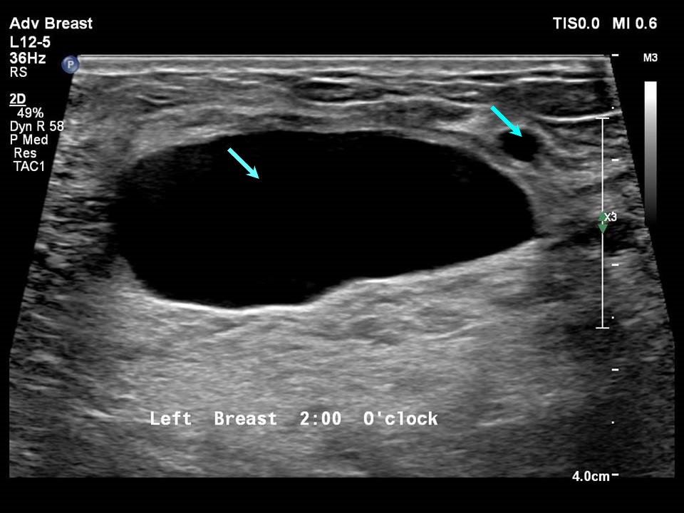
https www researchgate net publication 371137996 figure fig3 AS 11431281172713067 1688639457275 Axial section of MRI T2 sequence showing the cyst in hypersignal with a multivesicular png - Axial Section Of MRI T2 Sequence Showing The Cyst In Hypersignal With A Axial Section Of MRI T2 Sequence Showing The Cyst In Hypersignal With A Multivesicular https www researchgate net publication 365688591 figure fig2 AS 11431281099632629 1669280061644 S Detect Interpretation of a Cyst on Standard of Care and VSI B mode ultrasound images png - S Detect Interpretation Of A Cyst On Standard Of Care And VSI B Mode S Detect Interpretation Of A Cyst On Standard Of Care And VSI B Mode Ultrasound Images

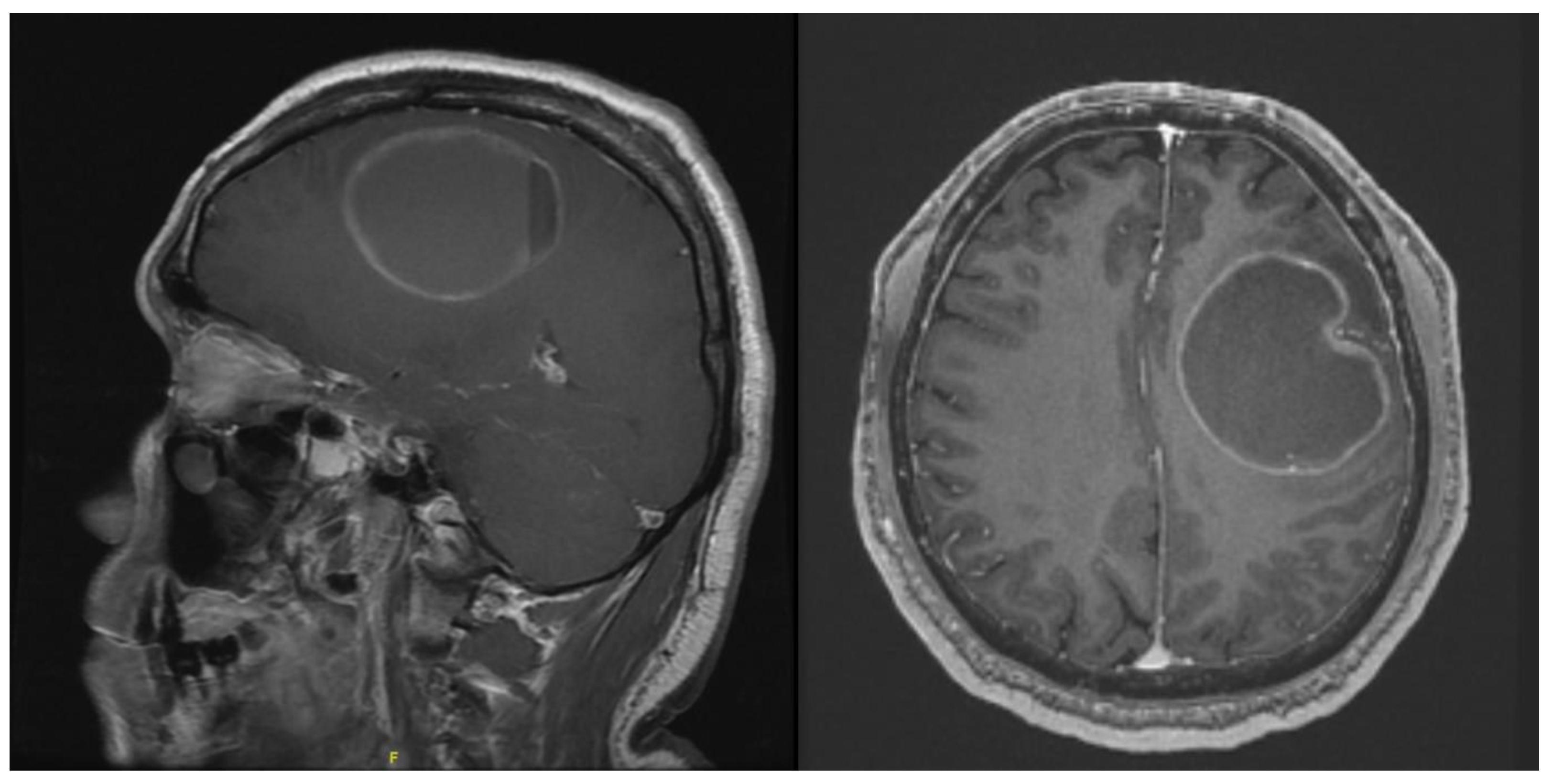
https www frontiersin org files Articles 932219 fneur 13 932219 HTML r1 image m fneur 13 932219 g001 jpg - Cat Scan Brain Tumor Fneur 13 932219 G001 https www researchgate net publication 367662499 figure fig2 AS 11431281116485261 1675255721168 MRI results at time of initial visit Arrow indicates cystic lesion A Axial Q320 jpg - MRI Results At Time Of Initial Visit Arrow Indicates Cystic Lesion MRI Results At Time Of Initial Visit Arrow Indicates Cystic Lesion A Axial Q320 https www researchgate net publication 304658452 figure fig1 AS 378975911202816 1467366306860 MRI T2 weighted cross section view showing the cyst in the anterolateral aspect of the png - MRI T2 Weighted Cross Section View Showing The Cyst In The MRI T2 Weighted Cross Section View Showing The Cyst In The Anterolateral Aspect Of The

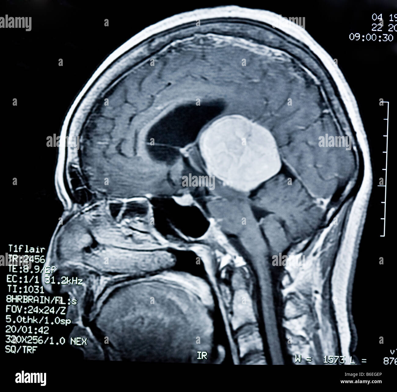
https www researchgate net publication 369287826 figure fig3 AS 11431281133302060 1680278724387 A Axial T1 post contrast MRI showing a 50 mm cystic mass with a solid intracystic jpg - A Axial T1 Post Contrast MRI Showing A 50 Mm Cystic Mass With A Solid A Axial T1 Post Contrast MRI Showing A 50 Mm Cystic Mass With A Solid Intracystic https www researchgate net publication 317024693 figure fig1 AS 868379757932545 1584049276450 MRI A multiple cystic tumor is recognized as a high intensity mass by T1 and T2 weighted png - MRI A Multiple Cystic Tumor Is Recognized As A High Intensity Mass By MRI A Multiple Cystic Tumor Is Recognized As A High Intensity Mass By T1 And T2 Weighted

https www researchgate net profile Seidu A Richard publication 338641291 figure fig2 AS 848370432237568 1579278681170 A C MRIs showing a cystic solid tumor MRI magnetic resonance imaging png - A C MRIs Showing A Cystic Solid Tumor MRI Magnetic Resonance A C MRIs Showing A Cystic Solid Tumor MRI Magnetic Resonance Imaging